Quantify skin health and treatment effects

Transform 15-second OCT scans into quantitative, actionable data
Quantify skin health
and treatment effects
VivoTools is an advanced software option for VivoSight Dx Pro, extending non-invasive OCT and D-OCT imaging beyond visual interpretation to deliver quantitative, repeatable measurements of skin structure and vascular function.
Designed for research, product development and claims substantiation, VivoTools enables faster, more consistent analysis—without the subjectivity, time and cost associated with biopsies or expert panel scoring.
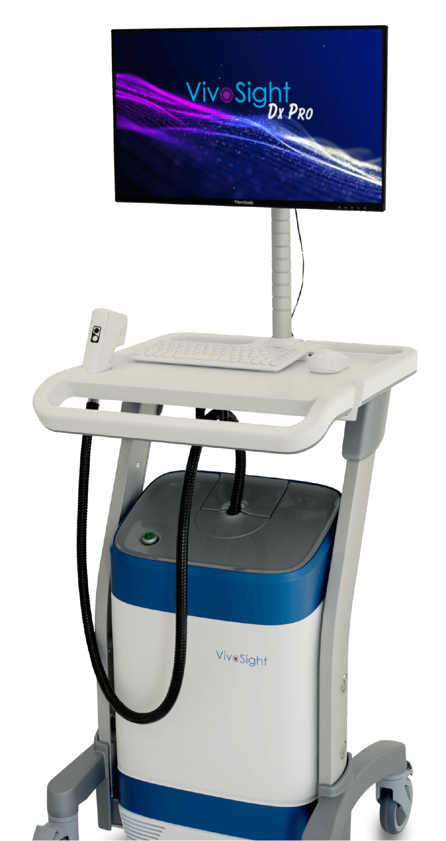
- Fully integrated with VivoSight Dx Pro for non-invasive OCT and D-OCT analysis.
- Automated extraction and quantification of key skin quality metrics, including changes in skin collagen content, reducing observer variability.
- Batch processing of scans supports high-throughput studies and maximises research productivity.
- Export images and numerical data for presentations, peer-reviewed publications and regulatory submissions.
- Collaborative, multi-seat capability enables consistent analysis across teams and sites.
- Highly cost effective – significantly reduces time and cost compared with biopsies, histology and expert panel evaluations.
Comprehensive quantitative skin assessment
Many conventional skin health assessments are subjective, qualitative and difficult to reproduce.
VivoTools enables a transition to objective, quantitative and repeatable measurement, using high-resolution, non-invasive OCT imaging.
Whether used by academic researchers, CROs, or cosmetics R&D and regulatory scientists, VivoTools supports more robust skin health monitoring, faster formulation development and stronger claims validation.
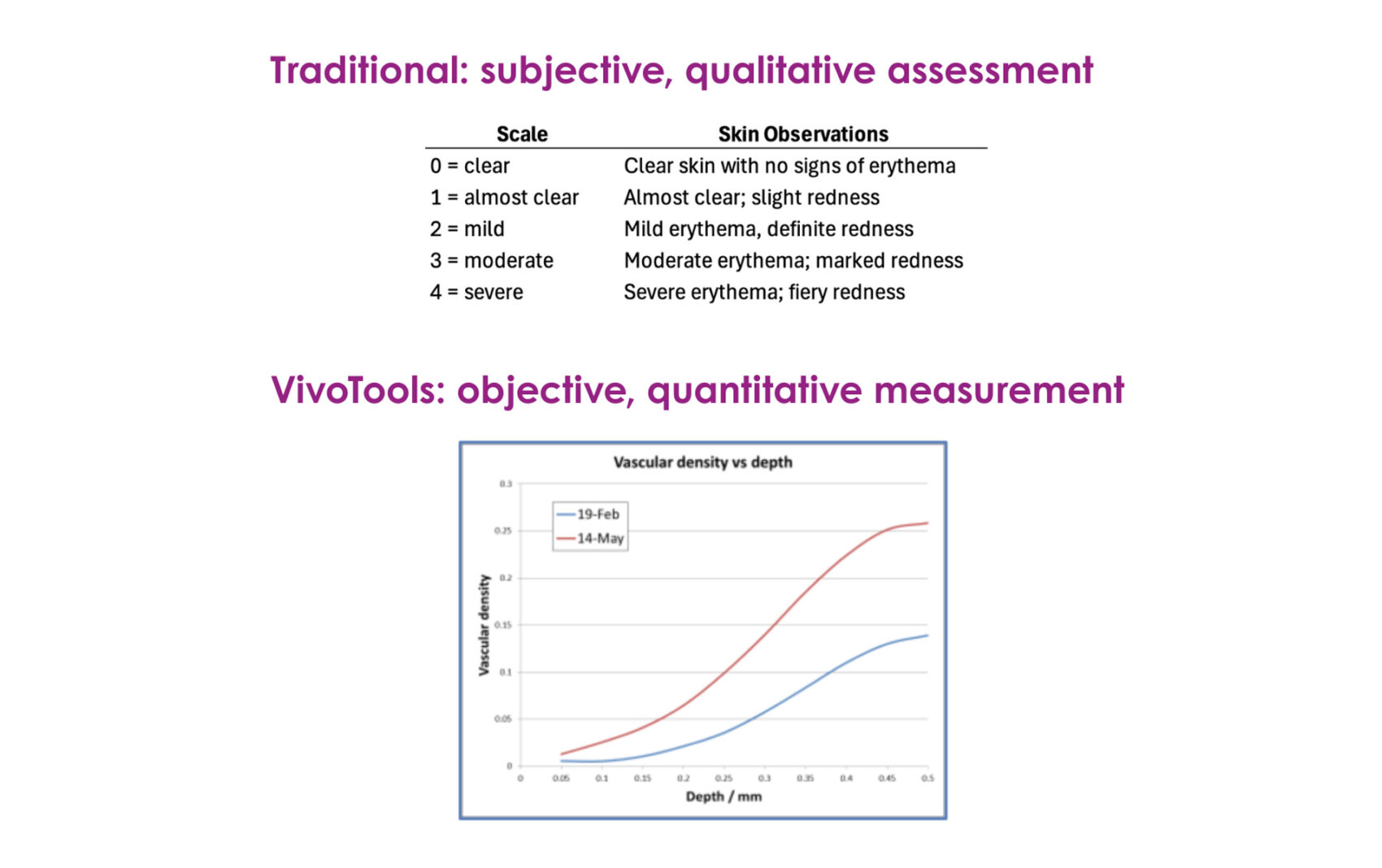
VivoTOOLs replaces a subjective erythema scale with quantification of vascular density
Taking just seconds to provide quantitative insight
VivoTools automatically extracts and quantifies the skin parameters that matter most — delivering objective, repeatable data from every OCT scan:
- Dermal collagen changes assessed via dermal brightness as a validated proxy marker
- Epidermal thickness
- Vessel diameter and vascular density by depth
- Skin surface roughness
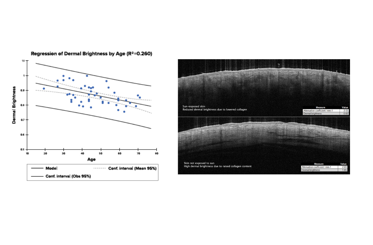
Assess Collagen changes through Dermal Brightness proxy
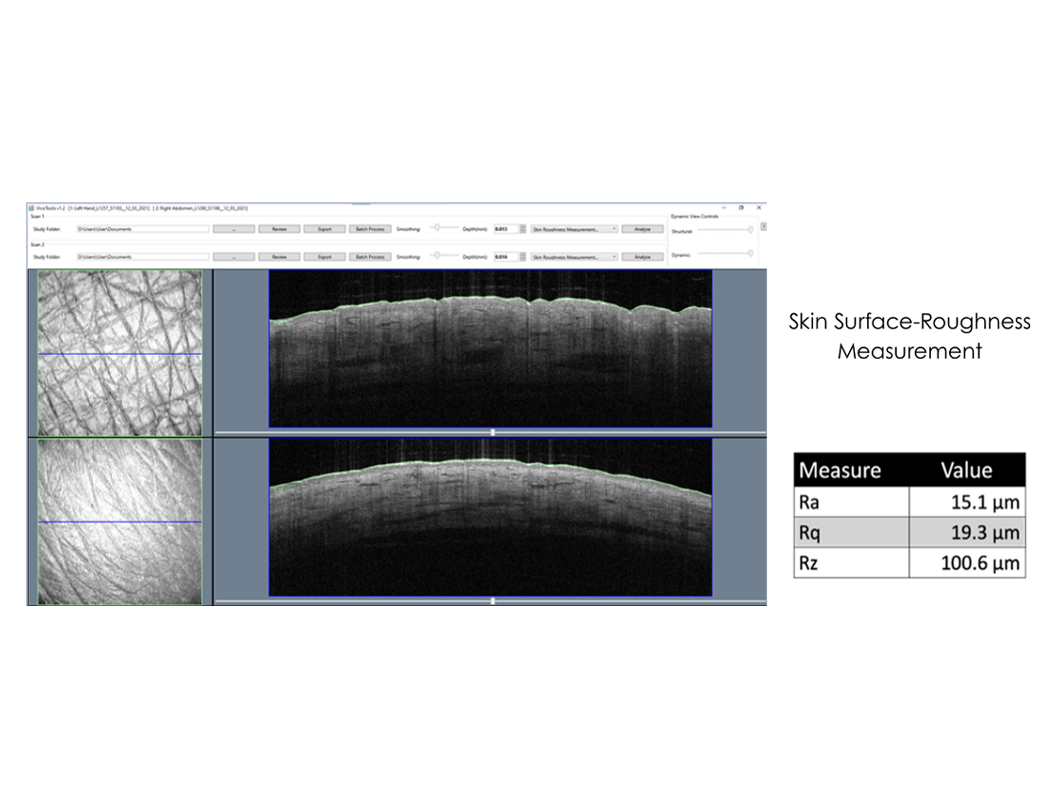
Quantify peak and average skin surface-roughness
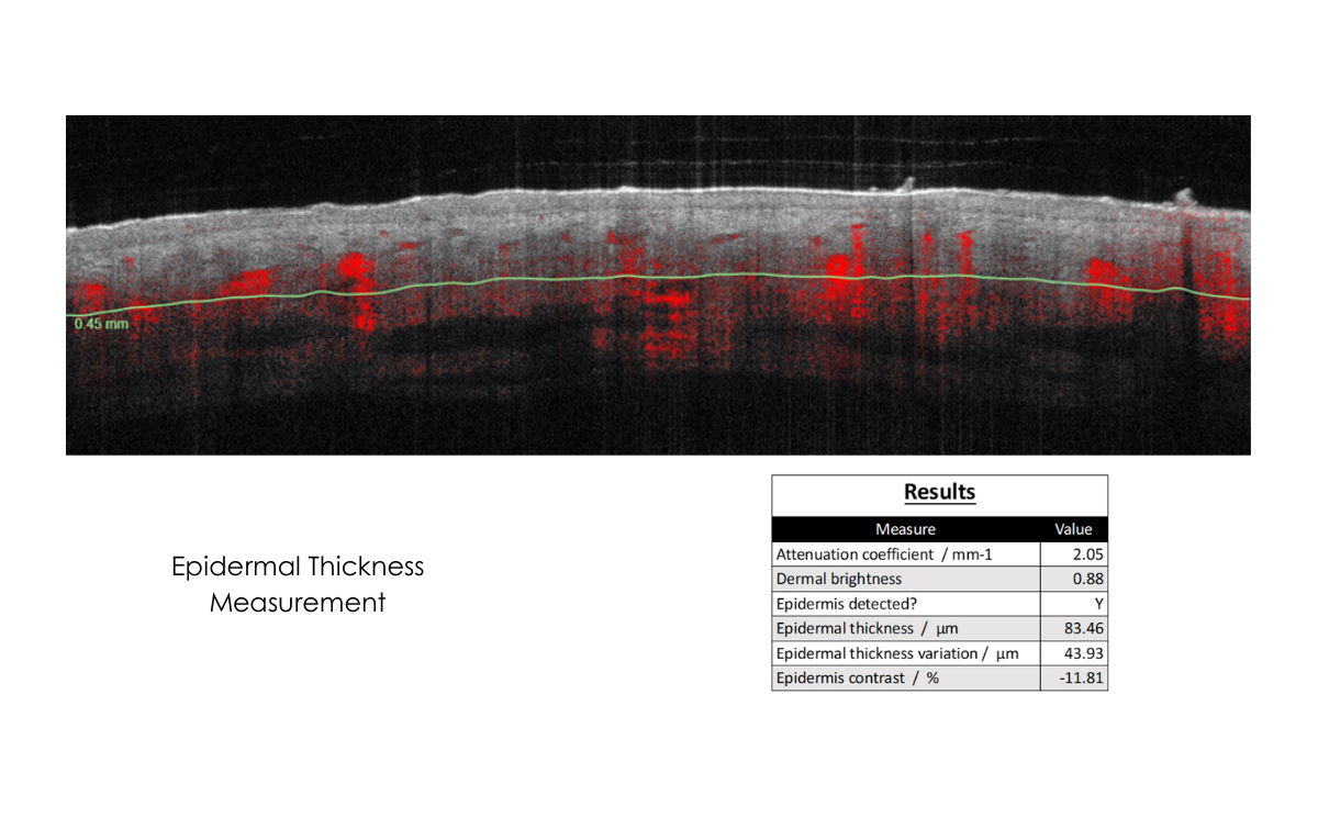
Measure Epidermal thickness
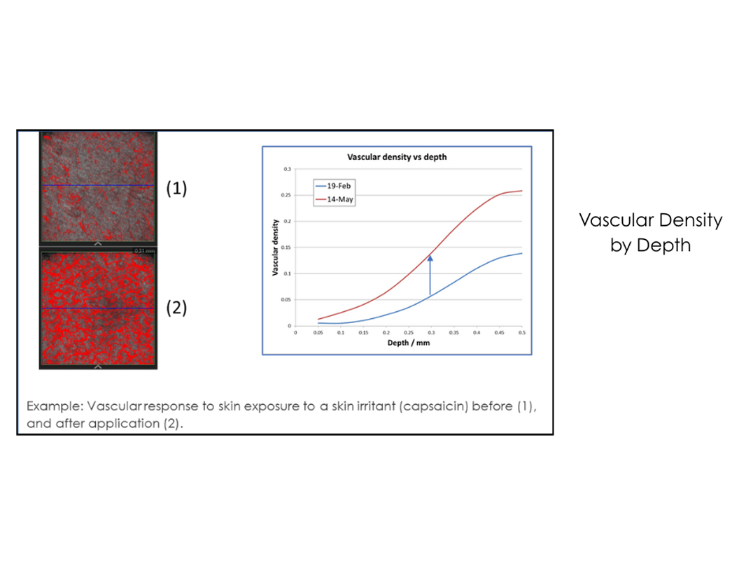
Quantify Vascular Density by depth
Clinically validated technology
VivoSight OCT technology has been cited in 800+ peer-reviewed publications
demonstrating its reliability and widespread adoption across dermatology, skin research and product development.
See VivoTools in Action
Discover how VivoTools can extend the capabilities of VivoSight Dx Pro.
Schedule your online demo today.